Computational Neuroscience

Foveated Retinotopy Improves Classification and Localization in CNNs
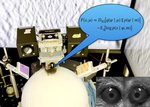
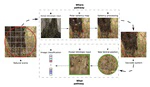
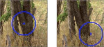
*Foveated Retinotopy in CNNs.* We represent Left an input image and how it is transformed by foveated retinotopy. We show below a representative reconstruction showing that it also acts as a cortical zoom on the image around the point of fixation. The transformed image is then fed to the ResNet deep learning architecture.
Foveated Retinotopy in CNNs. We represent Left an input image and how it is transformed by foveated retinotopy. We show below a representative reconstruction showing that it also acts as a cortical zoom on the image around the point of fixation. The transformed image is then fed to the ResNet deep learning architecture.

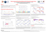
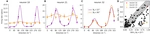
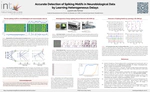
From falcons spotting prey to humans recognizing faces, the ability to rapidly process visual information depends on a foveated retinal organization that provides high-acuity central vision while preserving low-resolution peripheral vision. This organization is conserved along early visual pathways, yet remains under-explored in machine learning. Here, we examine the impact of embedding a foveated retinotopic transformation as a preprocessing layer on convolutional neural networks (CNNs) for image classification. By applying a log-polar mapping to off-the-shelf models and retraining them, we achieve comparable accuracy while improving robustness to scale and rotation. We demonstrate that this architecture is highly sensitive to shifts in the fixation point and that this sensitivity provides an effective proxy for defining saliency maps that facilitate object localization. Our results demonstrate that foveated retinotopy encodes prior geometric knowledge, providing a solution for visual searches and a meaningful classification robustness and localization trade-off. These findings provides a proof of concept in order to connect principles of biological vision with artificial networks, suggesting new, robust and efficient approaches for computer vision systems.

Foveated Retinotopy Improves Classification and Localization in CNNs
Foveal Retinotopy and Dual Pathways: A Computational Model for Active Visual Search

Abstract

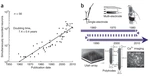
This thesis investigates visual search through the lens of the dual visual pathways found in biological systems : the ventral (“what”) pathway, involved in object recognition, and the dorsal (“where”) pathway, responsible for spatial localisation and saccadic planning. Drawing from both neuroscience and computer vision, we propose a computational framework that integrates deep convolutional neural networks (DCNNs) within a biologically inspired architecture grounded in foveal retinotopy. As a proof of concept, prior work has demonstrated that incorporating saccadic planning improves digit categorisation performance in a controlled environment. Building upon this foundation, the primary objective of this thesis is to extend the computational framework to natural images in more ecologically valid settings. Our contributions are as follows : (1) We introduce a novel framework for training and evaluating DCNNs using semantically grounded, task-specific labels ; (2) We bridge the gap between artificial models and biological substrates by emphasizing the role of foveal retinotopy in robust object categorisation and precise localisation ; (3) We disentangle the interplay between categorisation and localisation by proposing a novel “localisation-frame” dataset, aimed at guiding the design of a biologically plausible dorsal stream model ; and (4) We present an initial model of the dorsal pathway, leveraging the new dataset to develop interpretable and efficient active vision systems—where interpretability is achieved through modular and spatially structured representations, and efficiency is reflected in reduced computational cost during inference with saccade planning. Overall, this thesis extends the dual-stream computational paradigm for visual search, contributes tools for explainable active vision, and offers a platform to explore hypotheses about functional specialisation in the human visual cortex.